NEWS
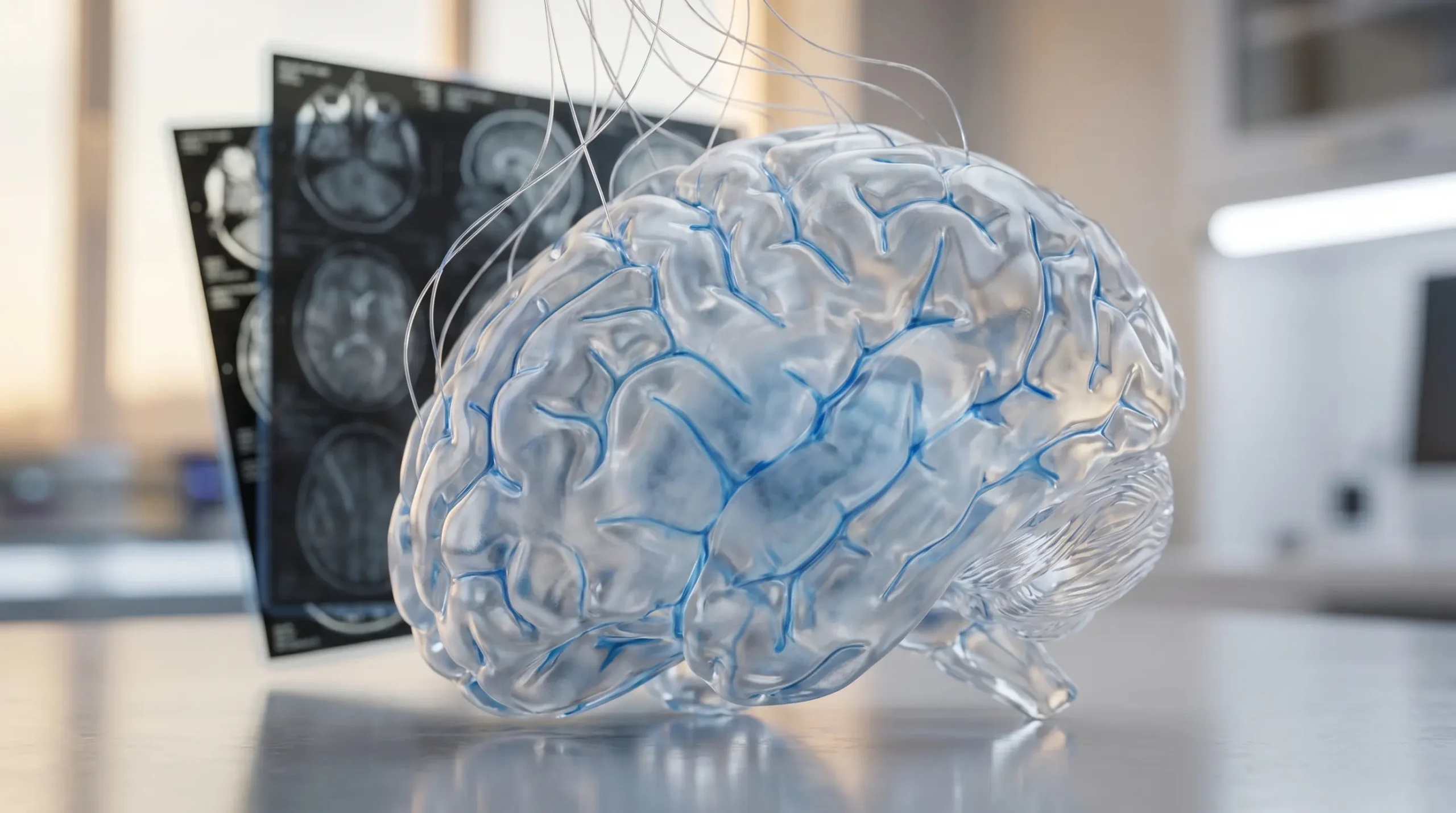
AI Brain Waste Study Maps the Brain’s Cleanup Speeds
An AI brain waste study published in the Science Advances MR-AIV study used magnetic resonance imaging (MRI, a scan that creates three dimensional pictures of internal tissue) and physics-informed neural networks to estimate how fast waterlike fluid moves through the brain’s glymphatic system. In mice, the method separated fast surface routes from a deep tissue trickle, a step toward non-surgical tests for clearance failures tied to Alzheimer’s disease and concussion.
For a field that has argued for more than a decade about whether the sleeping brain washes itself, the advance is practical: a speed map. The software narrows a stubborn measurement problem, giving researchers a way to infer flow that standard MRI can see only indirectly.
A Brain Cleanup Theory Gets a Speedometer
The glymphatic system is the brain’s waste-clearance route, a network in which cerebrospinal fluid (CSF, the clear liquid around the brain and spinal cord) exchanges with interstitial fluid inside tissue. Maiken Nedergaard, co-director of the University of Rochester Center for Translational Neuromedicine, and Jeffrey Iliff, then a researcher in her lab, helped put the system on the map in mice in 2012 through the University of Rochester glymphatic discovery report.
The idea caught on because it offered a physical answer to an old question: how does a high-energy organ without ordinary lymphatic vessels clear metabolic waste? Amyloid beta, one of the proteins linked to Alzheimer’s disease, made the question more urgent.
Douglas Kelley, professor of mechanical engineering at the University of Rochester, has worked on the plumbing problem from the physics side. Optical microscopes can track tiny particles with great detail near the brain surface, but they see only a small patch. MRI can view the whole brain, but the flow is so slow that the scanner does not directly return velocity.
That gap is where the new work sits. It asks less glamorous questions than a drug trial, but they are the questions clinics would need answered first: how fast does the fluid move, where does it slow down, and which tissue routes are doing most of the work?
Two Speeds, One Clearance Route
The team’s tool is called Magnetic Resonance Artificial Intelligence Velocimetry (MR-AIV, a framework that estimates velocity from MRI tracer movement while obeying fluid physics). The researchers used dynamic contrast-enhanced MRI (DCE-MRI, time-lapse MRI after an injected tracer) videos of dye spreading through mouse brains, then trained the model to infer velocity, pressure and tissue permeability.
The result was not one neat brainwide stream. It was a split pattern: fast advective flow around open spaces and perivascular routes, and much slower diffusion-driven transport through deep tissue.
- about 3 micrometers per second: the rapid advective flow scale reported for faster routes.
- about 0.1 micrometers per second: the slow diffusion-driven transport scale reported in the paper’s abstract.
- five wild-type mice: the in vivo animal data set used for the brainwide velocity maps.
A University of Rochester summary put the slower deep-brain route at about 50 times slower than the surface and open-region flow. The exact ratio matters less than the separation. If clearance fails in disease, the failure may come from a clogged highway, a sluggish sponge, or the exchange between the two.
The AI Is Doing Physics, Not Pattern Matching
Artificial intelligence in medicine often means pattern recognition: scan thousands of images, find a tumor, flag a risk. MR-AIV has a narrower job. It uses neural networks, but the model is constrained by equations that describe how tracers move through fluid and porous tissue.
That matters because the scanner records concentration over time, not flow speed. The hidden variables are reconstructed by making the model satisfy both the observed tracer movie and the rules of transport.
- Clean signal module: separates the tracer pattern from measurement noise before physics is applied.
- Pressure and permeability modules: estimate fields the MRI data cannot directly show.
- Transport equations: connect the changing tracer concentration to advection, diffusion and Darcy’s law, the rule commonly used for flow through porous material.
Juan Diego Toscano, a Brown University doctoral researcher, Yisen Guo, a University of Rochester computational scientist, and collaborators built the approach with Kelley, Kimberly Boster, University of Rochester assistant professor, Yuki Mori, University of Copenhagen associate professor, and George Karniadakis, Brown University professor of applied mathematics. The cast explains the method: neuroscience supplied the question, imaging supplied the data, and fluid mechanics supplied the guardrails.
Why Old Brain Imaging Hit a Wall
The study’s value is easier to see when the older tools are placed side by side. Each method sees part of the glymphatic system. None has been able to give a whole-brain, deep-tissue speed map without heavy inference.
| Method | What It Captures | Why It Falls Short | Where MR-AIV Helps |
|---|---|---|---|
| Two-photon microscopy | Particle motion near the brain surface | High detail, but a tiny field of view and invasive preparation | Extends inference beyond a small optical window |
| Dye front tracking | The advance of a tracer boundary | Can miss diffusion and out-of-plane movement | Uses the full tracer movie, not just the leading edge |
| Standard DCE-MRI | Whole-brain tracer concentration over time | Shows where tracer goes, not the slow velocity field | Adds physics-based velocity, pressure and permeability estimates |
| MR-AIV | Inferred brainwide transport fields | Depends on model assumptions and careful validation | Turns time-lapse MRI data into testable flow maps |
The table shows the narrow gain: MRI remains the measuring device; the AI adds a physics layer for variables the scanner leaves hidden. That makes the approach less flashy than a black-box diagnostic and more useful to a field still fighting over basic mechanics.
The Mouse Data Point Toward Humans, Cautiously
The mouse work is still far from a screening test. The Science Advances paper used preclinical imaging, a contrast agent delivered into the cisterna magna, and a small animal cohort. The models were trained on baseline measurements, not on a large disease population.
Still, the direction is clear in the University of Rochester MR-AIV announcement: compare healthy and sick brains, young and old brains, and eventually human brains. Kelley said the clinical applications become more important once researchers can measure waterlike fluid flow in and around people, especially for Alzheimer’s disease and concussion.
The hard part is the human scan problem. A clinical method would need repeatable measurements, safe and acceptable tracer protocols, and proof that a slow-flow pattern predicts something useful. A beautiful map in a mouse brain is a beginning, not a biomarker.
That caution should not bury the advance. Before doctors can ask whether a patient’s brain is clearing waste poorly, researchers need a normal range, a disease range and a way to compare the two. MR-AIV is aimed at that missing ruler.
Sleep Science Still Has a Debate to Settle
The glymphatic field also carries live controversy. In 2024, the Nature Neuroscience sleep clearance study from researchers at Imperial College London reported that brain clearance in male mice was reduced during sleep and anesthesia, challenging a leading interpretation of earlier work.
Other human and animal studies point in a different direction. A 2026 Nature Communications human glymphatic study reported a randomized crossover trial with 39 participants and found evidence consistent with sleep-active clearance of amyloid beta and tau from brain to plasma. Higher morning plasma biomarkers can sound alarming, but in that model it may reflect proteins moving out of the brain into blood.
Five mice cannot settle that debate. The method gives future teams a common measure: where fluid moves, how fast it moves, and which pathways change under sleep, wakefulness, aging or disease. That is a better argument than dueling metaphors about brain washing.
Clinics Need Baselines Before Diagnoses
The research program behind the paper is larger than one algorithm. The NIH-backed U19 fluid and solute clearance project brings together mouse and human work on how neural activity, blood volume and CSF movement interact during sleep. MR-AIV gives that program a quantitative imaging tool.
Alzheimer’s disease is the obvious target, but concussion may be the cleaner early test case. After a head injury, doctors already need better ways to know when brain physiology has recovered. A fluid-circulation measure could add information that ordinary structural imaging misses, provided future studies show the signal is reliable.
If the method translates to human scans, the field gains a ruler for a system that has mostly been inferred. If it does not, the mouse maps still set a sharper benchmark for the next experiment.
-
 NEWS10 years ago
Samsung Releases Galaxy Note7 TV Ad as Reddit AMA Leaks Specs
-
 NEWS10 years ago
Android 7.0 Nougat Rolls Out To Nexus Devices With New Emoji, Features
-
 FINANCE8 years ago
Cardano Price Surges as ADA Enters the Crypto Top Ten List
-
 NEWS10 years ago
Pre-Order the First Camera Made for Facebook Live Streaming Video
-
 FINANCE8 years ago
RChain Price Jumps Nearly 150% to a New All-Time High of $2.03
-
 FINANCE10 months ago
Binance Suspends Trading and Withdrawals for a System Upgrade
-
 NEWS10 years ago
Google Play App Icons Get Fresh New Look: See the Latest Design Update
-
 NEWS10 years ago
Google Doodle Go Bananas Fruit Games Live On Mobile For Two Weeks