Stat3 proteins are essential for sending signals inside our cells, controlling everything from immune responses to cell growth. For Stat3 to work, two of these proteins must pair up in a process called dimerization. This connection happens at a specific location called the dimerization interface, which relies on particular amino acids to form a stable bond. Understanding these key amino acids helps scientists learn how to control Stat3 activity, which is crucial for developing treatments for diseases like cancer.
Understanding the Structure of a Stat3 Protein
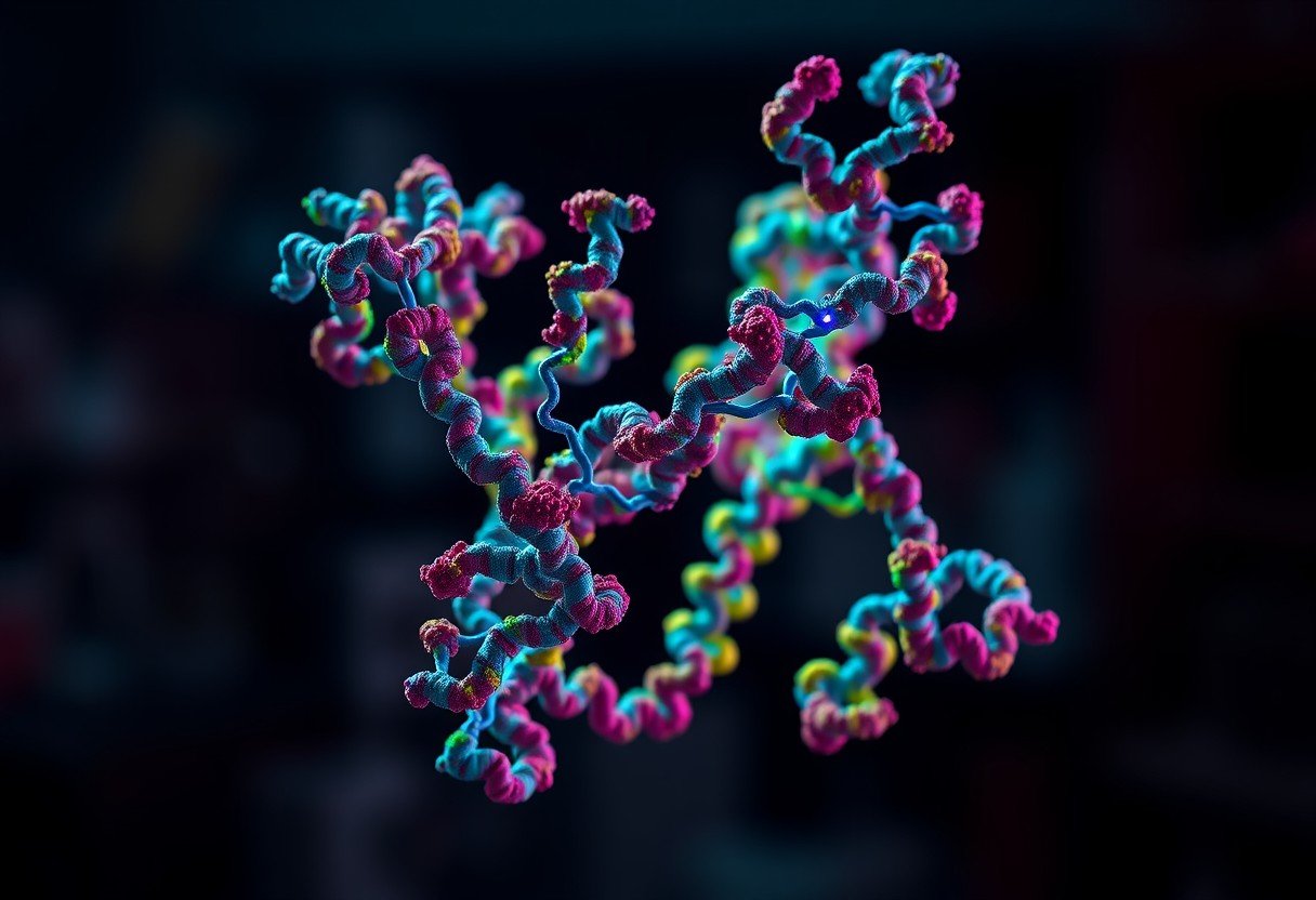
To grasp how Stat3 proteins pair up, it’s helpful to think of them as having different sections, or domains, each with a specific job. A Stat3 protein is made up of an N-terminal domain, a coiled-coil region, a DNA-binding domain, and a C-terminal domain. Each part plays a role in its overall function.
The coiled-coil region is especially important for dimerization. It acts like a magnet, drawing two Stat3 proteins together to form either a homodimer (two identical Stat3 proteins) or a heterodimer (with a different protein). This pairing is a necessary step before the protein can travel to the cell’s nucleus and turn genes on or off.
Once dimerized, the protein changes shape, revealing its DNA-binding domain. This allows the dimer to attach to specific DNA sequences and regulate gene expression. Therefore, the structural integrity of each domain, especially the coiled-coil region, is vital for Stat3’s ability to function correctly.
The Crucial Role of Different Amino Acid Types
The dimerization of Stat3 proteins is not random; it depends heavily on the specific amino acids present at the interface. These amino acids create the forces that hold the two proteins together, ensuring the dimer is stable enough to perform its job. The interactions are driven by two main types of amino acids: hydrophobic and charged/polar.
Hydrophobic amino acids, which dislike water, play a huge part in stabilizing the dimer. They cluster together at the core of the interface, pushing water away and creating a tight, secure bond. This hydrophobic interaction is a primary driving force for the formation of many protein complexes.
On the other hand, charged and polar amino acids contribute to the specificity and stability of the connection through hydrogen bonds and electrostatic interactions. These forces act like tiny magnets, ensuring the two Stat3 proteins align perfectly. This combination of hydrophobic and electrostatic forces creates a robust and highly specific dimerization interface.
Key Amino Acids Driving the Dimerization Process
While many amino acids are involved, some are more frequently found at the Stat3 dimerization interface due to their unique properties. Identifying these key players gives researchers valuable clues for understanding and manipulating Stat3 activity.
Hydrophobic residues are the stars of the show when it comes to stability. They form the core of the interface.
- Leucine and Isoleucine: These are commonly found because their bulky, nonpolar side chains are excellent for creating strong hydrophobic interactions.
- Phenylalanine and Methionine: These also contribute to the water-excluding core, further strengthening the dimer.
Polar and charged residues provide the necessary specificity and fine-tuning. They form hydrogen bonds and salt bridges that lock the dimer into its active conformation. For instance, serine and threonine can form critical hydrogen bonds, while charged residues like arginine and glutamic acid can create powerful electrostatic attractions.
| Amino Acid Type | Common Examples | Primary Role in Dimerization |
|---|---|---|
| Hydrophobic (Nonpolar) | Leucine, Isoleucine, Phenylalanine | Forms a stable core and drives dimer formation. |
| Polar | Serine, Threonine | Participates in hydrogen bonding for stability. |
| Charged | Arginine, Glutamic Acid | Creates electrostatic attractions for specificity. |
How Scientists Identify these Interface Amino Acids
Pinpointing the exact amino acids at the dimerization interface requires a combination of sophisticated laboratory techniques and powerful computational tools. These methods allow scientists to visualize the protein structure and test the function of individual residues.
Experimental techniques provide a direct look at the protein’s three-dimensional shape. X-ray crystallography and Nuclear Magnetic Resonance (NMR) spectroscopy are two of the most powerful methods used to determine the precise structure of the Stat3 dimer. These techniques reveal which amino acids are in close contact at the interface.
In addition to structural studies, researchers use bioinformatics to predict which regions are likely to be involved in dimerization. By comparing Stat3 protein sequences across different species, scientists can identify conserved amino acids that are probably essential for function. Molecular dynamics simulations can then be used to model how changes to these amino acids might affect the stability of the dimer, complementing experimental findings.
Targeting the Dimerization Interface for Cancer Therapy
Because Stat3 activation is often linked to the growth and survival of cancer cells, its dimerization interface has become an attractive target for new therapies. Many cancers show constant, or aberrant, Stat3 activity, which helps tumors grow, resist treatment, and spread. By preventing Stat3 from dimerizing, it’s possible to shut down this pro-cancer signaling pathway.
Researchers are actively developing small-molecule inhibitors designed to block the Stat3 dimerization interface. These drugs work by physically getting in the way, preventing the two Stat3 proteins from coming together. Without the ability to form a dimer, Stat3 cannot travel to the nucleus to activate the genes that fuel cancer progression.
This targeted approach holds great promise for treating various malignancies where Stat3 plays a key role. Combining these Stat3 inhibitors with traditional therapies could also make treatments more effective, especially for cancers that have become resistant to other drugs. This represents a significant step forward in developing more precise and effective cancer treatments.
Frequently Asked Questions about Stat3 Dimerization
What is the main function of the Stat3 protein?
Stat3 is a transcription factor, meaning its main job is to regulate gene expression. It responds to signals from outside the cell, travels to the nucleus, and turns specific genes on or off to control processes like cell growth, survival, and immune response.
Why is dimerization necessary for Stat3 to work?
Dimerization is a required activation step for Stat3. Forming a dimer exposes the protein’s DNA-binding domains and allows it to translocate into the cell nucleus, where it can bind to DNA and carry out its function. A single Stat3 monomer is inactive.
Which amino acids are most common at the dimerization interface?
The interface typically contains a mix of amino acids. Hydrophobic residues like leucine and isoleucine are very common because they provide stability, while charged residues such as arginine and glutamic acid help with the specific alignment of the two proteins.
What happens if an amino acid at the interface is mutated?
A mutation at the dimerization interface can disrupt the stable bond between the two Stat3 proteins. This can impair or completely block Stat3’s function, leading to faulty cell signaling, which can contribute to various diseases, including cancer and autoimmune disorders.
How does phosphorylation affect Stat3 dimerization?
Phosphorylation, the addition of a phosphate group, is a critical post-translational modification for Stat3. The phosphorylation of a specific tyrosine residue allows two Stat3 monomers to recognize each other and form a stable dimer, effectively switching the protein on.


Leave a Comment